Pharmaceutical Methods
Publishing Quality Research & Reviews
Pharmaceutical Methods
Publishing Quality Research & Reviews
Perspective - (2022) Volume 13, Issue 2
Received: May 20, 2022, Manuscript No. PHMETHODS-22-66360; Editor assigned: May 23, 2022, Pre QC No. PHMETHODS-22-66360 (PQ); Reviewed: Jun 06, 2022, QC No. PHMETHODS-22-66360; Revised: Jun 13, 2022, Manuscript No. PHMETHODS-22-66360 (R); Published: Jun 21, 2022, DOI: 10.35248/2229-4708.22.13.230
Fluorescence Correlation Spectroscopy (FCS) is a statistical analysis, by time correlation, of stationary fluctuations of the fluorescence intensity. The analysis provides kinetic parameters of the physical processes underlying the fluctuations. One of the interesting claims of this is an analysis of the concentration fluctuations of fluorescent particles (molecules) in solution. In this application, the fluorescence emitted from a very tiny space in solution enclosing a small number of fluorescent particles (molecules) is observed. The fluorescence intensity is unstable due to Brownian motion of the particles.
Fluorescence Correlation Spectroscopy (FCS) has been used to study the guest host complexes involving γ-cyclodextrin (γ- CD) as a host and coumarin dyes (C480 and C153) as guest. It is shown that C480 forms a nearly spherical 1:1 complex with -CD. C153 on the other hand, forms a large nano-tube aggregate with -CD. This aggregate is non-spherical and resemble a rod or needle. For this system the standard Stokes-Einstein model is not valid. It determined the length of the γ-CD:C153 nanotube aggregate to be ~ 770 nm. This is ~ 480 times larger than the length of a 1:1 γ-CD:C480 complex (~ 1.6 nm) and ~ 950 times that of a γ-CD. This implies that 950 γ-CD units are non-covalently attached in the γ-CD:C153 aggregate. Binding constants (Kb) of both the dyes to γ-CD were obtained from the fluctuation in fluorescence intensity. The rate of association and dissociation are obtained from the inverse of Եoff and Եon, respectively. The binding constant for the 1:1 γ-CD:C480 complex is ~ 103 M-1 and that for -CD:C153 aggregate is ~ 105 M-1 . The Burst Integrated Fluorescence Lifetime (BIFL) histogram of the nanotube aggregates reveals the presence of three distinct lifetime 1.8 ns (18%), 2.8 ns (69%), 3.2 ns (13%). These three lifetimes correspond to C153 present in bulk water and at the end and middle of the nanotube aggregate, respectively. The lifetime of C480 in the 1:1 γCD:C480 complex is found to be 3.7 ns.
The effect of two Room-Temperature Ionic Liquids (RTILs) on the diffusion of three fluorescent dyes in the gel phase of a triblock copolymer, (PEO)20-(PPO)70-(PEO)20 (Pluronic P123) was studied by Fluorescence Correlation Spectroscopy (FCS). We used three dyes, 4-(dicyanomethylene)-2-methyl- 6-(4-dimethylaminostyryl)-4H-pyran(DCM), coumarin 480(C480), and coumarin 343(C343). By Field-Emission Scanning Electron Microscopy (FESEM), it was observed that the macroscopic structure of the P123 gel remains unaffected upon addition of RTIL. In the absence of RTIL, the diffusion coefficient (Dt) of the hydrophobic dye DCM (1 m2s-1 at the core) is smaller than that of the other two hydrophilic dyes (7 m2 s-1 for C480 and C343). On addition of RTIL, the Dt values of all of the dyes increase, indicating a decrease in local viscosity (eff). The eff of the core of the RTIL-P123 gel estimated from the Dt of DCM is lower than that of both the P123 gel (at the core=90 cP) and RTIL (=110 cP). It is shown that the RTIL affects the structure of the gel by modifying the size of the micellar aggregates and by penetrating the core.
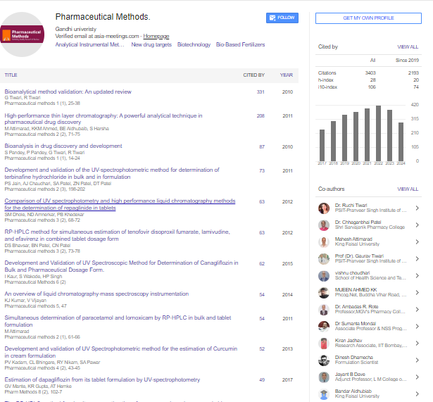
Pharmaceutical Methods received 3403 citations as per google scholar report